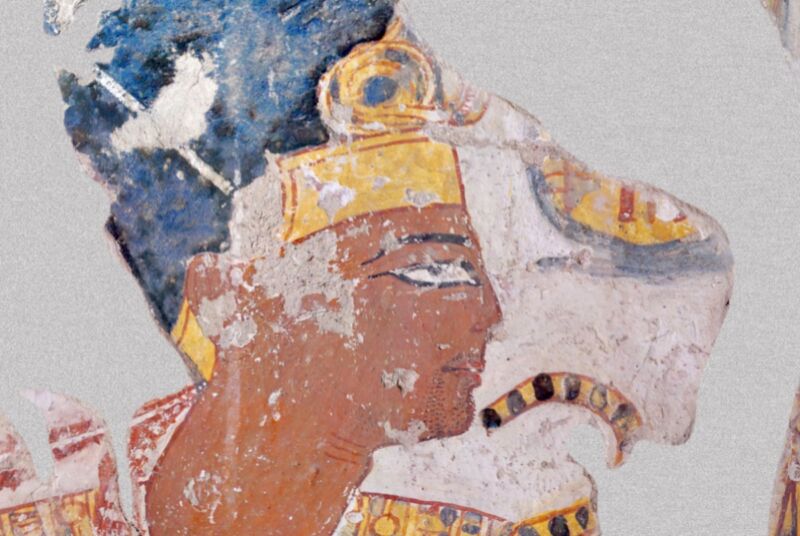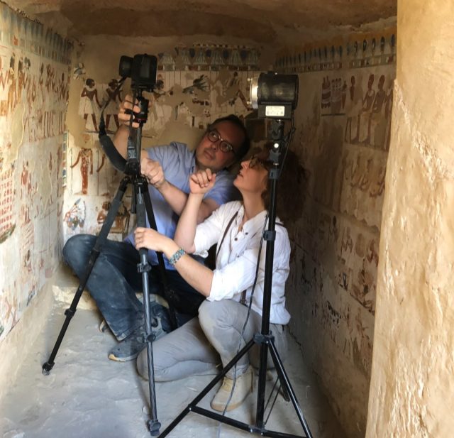
Researchers have discovered evidence of earlier versions of two ancient Egyptian paintings located in tomb chapels in the Theban Necropolis, according to a new paper published in the journal PLoS ONE. In one, there is a ghostly third hand partially hidden under a white overprinted layer; the other has adjustments to the crown and other royal items in a portrait of Ramesses II. These discoveries were made with a portable macro-X-ray fluorescence (MA-XRF) imaging device that enabled the researchers to analyze the paintings on site, with no need for taking physical samples.
X-rays are a well-established tool to help analyze and restore valuable paintings because the rays’ higher frequency means they pass right through paintings without harming them. X-ray imaging can reveal anything that has been painted over a canvas or where the artist may have altered his (or her) original vision. For instance, Vermeer’s Girl Reading a Letter at an Open Window was first subjected to X-ray analysis in 1979 and revealed the image of a Cupid lurking under the overpainting. In 2020, a team of Dutch and French scientists used high-energy X-rays to unlock Rembrandt’s secret recipe for his famous impasto technique, believed to be lost to history.
In 2021, scientists used macro-X-ray fluorescence imaging to map out the distribution of elements in the paint pigments of Jean-Louis David’s famous portrait of 17th-century chemist Antoine Lavoisier and his wife Marie-Anne—including the paint used below the surface—to create detailed elemental maps for further study. Earlier this year, scientists used X-ray powder diffraction mapping and synchrotron micro X-ray analysis to study Rembrandt van Rijn‘s 1642 masterpiece The Night Watch, and found rare traces of a compound called lead formate.
In 2022, Dutch and Belgian scientists used X-ray imaging techniques to examine the elemental distribution of the various pigments in a yellow rose prominently featured in 17th-century painter Abraham Mignon’s Still Life with Flowers and a Watch to glean insight into why it had become flattened and monochrome, particularly compared to the other blooms featured in the painting.

That same year, conservationists were conducting an X-ray analysis of Vincent van Gogh’s Head of a Peasant Woman and discovered a hidden self-portrait on the back of the canvas. Nor was this the first time a Van Gogh painting has been subjected to X-ray analysis. Back in 2008, European scientists used synchrotron radiation to reconstruct the hidden portrait of a peasant woman painted by Van Gogh.
MA-XRF imaging in particular has previously been used (in combination with hyper spectral imaging) to study a Greco-Roman painted funerary portrait from the second century CE, as well as determining the remnants of color on marble at Delphi in Greece. Now the portable MA-XRF imaging device developed for one of the Delphi studies is being applied to the study of ancient Egyptian paintings, part of a broader project investigating tomb chapels in the Theban Necropolis. Many tombs for high-ranking officials featured chapels whose walls were decorated with painted murals depicting the deceased in life as well as their future in the netherworld.
It is known that the ancient Egyptians had a highly formalized (and easily recognizable) painting style, and there has been considerable interest in gaining insights into the specific pigments and painting techniques employed to create the tomb murals. Scholars have generally assumed that the process began by sketching a preliminary outline in red ochre on a smooth plastered wall, followed by applying a white or colored background. Other colors were then applied in layers using various pigment mixtures, and the final outline was drawn primarily with red ochre. Any spillover paint was painted over with opaque white.

